Clinically, the term "neoplasm" means a local overgrowth of any tissue of the body. On the skin, they are represented by primary and secondary tumors, nevi and hemoderma.
In dermatological practice, tumors are divided into benign and malignant. A detailed photo and a detailed description of each of them will be given below.
Why do they arise
The study of skin neoplasms is still ongoing. The exact reasons for their occurrence have not been established, but scientists put forward several theories on this score.
Provoking factors can be:
- burdened heredity (presence of neoplasms in relatives);
- individual characteristics of a person (light skin and hair, old age);
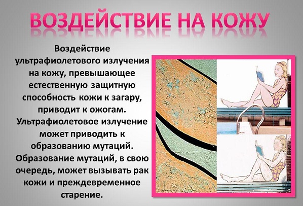
- exposure to ultraviolet, radiation and x-ray radiation;

- viral infection;
- prolonged trauma to the skin;
- chronic exposure to the skin of chemical carcinogens (nitrosoamines, benzopyrene, aromatic amines, etc.);
- insect bites;
- metastatic processes in the presence of an oncological process in the body;
- violation of the trophism of the skin, hence the chronic skin ulcers;
- a weakened immune system (due to immunosuppressive therapy, HIV infection, etc.).
Types of skin neoplasms
Neoplasms on the skin by their origin can be divided into primary (those that are formed from the skin tissue itself) and secondary (those that metastasize into the dermis and epidermis from foci of other localization). The latter also includes hemoderma. They arise due to the pathological proliferation of malignant cells of the hematopoietic system.
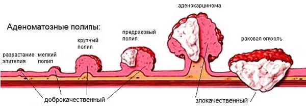
There is a division of neoplasms into benign, precancerous (precancerous) and malignant (cancer itself). This classification allows you to determine the method of treatment and life prognosis for the patient.
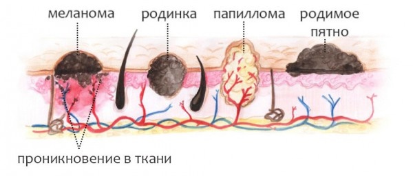
Nevi should be distinguished from skin tumors. These are benign neoplasms that belong to skin malformations.
Malignant neoplasms
Neoplasms on the skin of a malignant nature in the Russian Federation in the structure of oncological morbidity today amount to 9.8% and 13.7% in men and women, respectively. Persons living in areas with high photoinsolation and having fair skin are especially susceptible to the disease. The number of new cases of skin cancer has increased by a third over the past ten years.
Malignant skin tumors include:
- basal cell carcinoma;
- Kaposi's sarcoma;
- liposarcoma;
- squamous cell carcinoma;
- melanoma, etc.
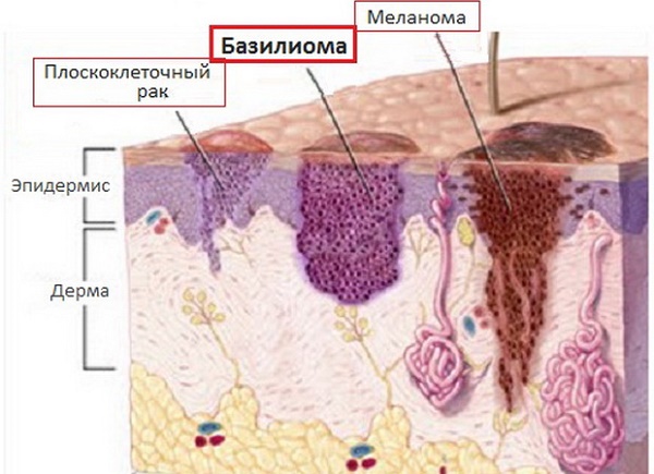
Basalioma
One of the most common epithelial skin tumors. It is formed from atypical cells of the basal layer of the epidermis, from where it got its name. The tumor is characterized by long-term progression, peripheral growth, during which the destruction of the surrounding tissues occurs. Basal cell carcinoma is not prone to metastasis.
This pathology develops mainly in the elderly and old people, localized mainly on the face, neck and head (its scalp). Sometimes basal cell carcinoma is referred to as precancer. under the influence of some factors, it degenerates into metatypical cancer.
The first manifestation of an emerging tumor is a dense hemispherical nodule that does not rise above the skin. Its color usually coincides with skin color or differs slightly (slight pink tint).
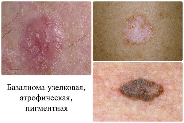
At the initial stage, the patient does not complain. Over the course of several years, the papule grows, reaching 1 or 2 cm in diameter. Its center gradually collapses, bleeds and crusts.
Under the latter, erosion or an ulcer with a narrow ridge along the edges is found, which over time scar and grows along the periphery.
Basalioma reaches a size of 10 or more centimeters. The once pink papule turns either into a flat plaque with scaling, or a node that rises noticeably above the surface of the skin, or a deep ulcer that destroys the underlying tissue (up to the bone).
Liposarcoma
This is a neoplasm on the skin of fat cells of mesenchymal origin. In the photos below, you can see what size these tumors reach. Descriptions in clinical reference books speak of liposarcoma as a mass that tends to appear on the buttocks, thighs, and retroperitoneal tissue. It is more common in men over 40.
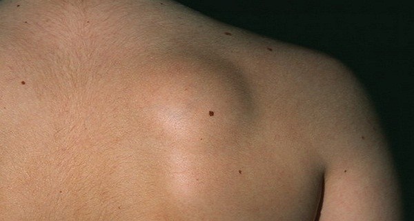
Initially, a swelling appears, then a node. There are no subjective sensations yet. On palpation, the nodule is dense, elastic, mobile.
Subsequently, the tumor grows, turns red, and inflammatory processes begin. Large liposarcoma can compress the nerves and blood vessels and even grow in them, causing tissue trophic disorders and pain.
Kaposi's sarcoma
This is a systemic multifocal disease of vascular origin with a predominant lesion of the skin, lymphatic system and internal organs. It refers to tumors of an endothelial nature and develops mainly in individuals with severe immunosuppression.
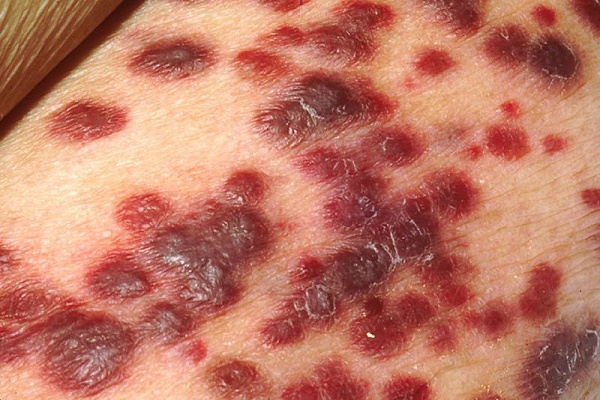
By morphology, skin foci of sarcoma are quite diverse. They come in the form of spots, nodules, infiltrative plaques, etc.
There are several types of sarcoma:
- Classic (European).
- Endemic (African).
- Epidemic (with HIV).
- Immunosuppressive (immunodeficiency caused by medications and therapeutic manipulations).
The first type is observed in the elderly and old people, has a favorable course. Elements grow for a long time, tens of years in the proximal direction and do not give the patient unpleasant sensations. The formations are most often localized on the lower extremities, are cyanotic-red spots up to 5 cm in diameter with smooth edges, resemble hematomas.
In the process of growth, they are transformed into nodules, merge. Large nodes darken and ultimately ulcerate. Edema occurs along the edges of the elements, caused by stagnation of lymph in the lymphatic system.
The African type is difficult, affecting the young. A fulminant course of the disease is often observed. African Kaposi's sarcoma manifests itself in several types of formations - from nodes to lymphadenopathy.
The most malignant type of elements of this type of sarcoma is considered "flowering" (growth in the form of vegetation - in appearance it resembles a cauliflower). It is characterized by deep lesions of the dermis, subcutaneous tissue and underlying tissues down to the bone.
With HIV infection, a tumor can be localized literally anywhere in the body, even affecting internal organs. The most common sites are the mouth, stomach and duodenum. The current is heavy. The immunosuppressive type is similar in its manifestation to the HIV-associated one.
Squamous cell carcinoma
Malignant tumor from the epithelium. It is formed from atypical keratinocytes that proliferate randomly.The process begins in the epidermis, gradually moving into deeper layers. The tumor is characterized by a tendency to metastatic process.
Squamous cell carcinoma occurs 10 times less often than basal cell carcinoma. They are more often affected by white-skinned men, whose place of residence is a sunny warm climate.
The localization of spinocellular epithelioma is different. The most favorite place for the formation of squamous cell carcinoma is the border of the transition of the mucous membrane into the skin. These areas include the lips and genitals.
At the initial stage of cancer development, an infiltrate with a towering hyperkeratotic (rough) surface occurs. The color of the formation is usually gray or yellow-brown.
Complaints at first, as with basal cell carcinoma, are absent. In the process of growth, the tumor can reach sizes up to 1 cm.At this moment, a dense knot is already felt, and continues to grow. Eventually, the carcinoma approaches the size of a walnut.
The tumor grows in two directions - above or deep into the tissues. The latter is usually accompanied by the formation of an ulcer, which affects not only the dermis and epidermis, but also reaches the bone and muscle tissue.
An ulcer in squamous cell carcinoma does not heal. The patient suffers from excruciating pain in the place of its formation. In the future, a violation of general well-being and infectious complications associated with immunosuppressive processes accompanying any oncopathology join the complaints.
Melanoma
This is a tumor of neuroectodermal origin. It consists of malignant melanocytes. The main provoking factor is considered to be UV radiation.
Melanoma develops from both a pre-existing nevus (birthmark) and clear skin.
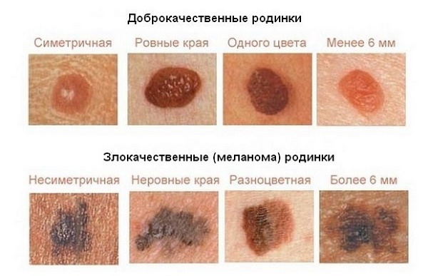
Signs of malignancy include:
- asymmetry;
- fuzzy edges;
- uneven color;
- diameter over 6 mm;
- the evolution of the age spot (any changes in the mole - sudden growth, discoloration, etc.) is the most typical sign!

 Don't miss the most popular column article: Amaranth oil - properties and application in cosmetology, reviews, the price of the product.
Benign neoplasms
Neoplasms on the skin belonging to the category of benign, as seen in the photo and by their description, are not prone to rapid growth, metastasis and relapse after removal.
These tumors include:
- atheroma;
- hemangioma;
- lymphangioma;
- warts;
- moles (nevi);
- fibroids, etc.
| View | Description |
| Atheroma | The defeat of the sebaceous gland in the form of cystic growth. The place of localization is the face. A raised formation, indistinguishable from normal skin in color, is tightly soldered to it at one point. The contours are clear. Palpation of atheroma is painless. |
| Hemangioma | Neoplasm of vascular genesis, refers to childhood tumors. It can be either an independent disease or a manifestation of another pathology. The course depends on the age of the patient, location, size and depth to which the hemangioma has grown. Frequent placement - head, face, neck, but other localization is also possible. The first manifestation is a red nodule (papule) up to 5 mm in diameter. From complaints - bleeding when accidentally touched, sometimes - dysfunction of the organ where the tumor is located. |
| Lymphangioma | There are two possible development options - congenital defect or consequences of impaired lymph flow. The tumor can be located anywhere, but more often it is the oral cavity, neck, upper limbs. Capillary lymphangiomas are multiple vesicles with a yellowish transparent liquid inside. The same tumors that have arisen as a result of impaired lymph circulation look like plaques or spots, the diameter of which is slowly increasing. |
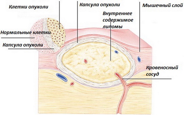
| Lipoma | Tumor of adipose tissue. Most often they have a nodal shape. The course is asymptomatic.It is a painless pale pink neoplasm on the pedicle. Doughy consistency. The borders are fuzzy. |
| Seborrheic warts | Also called "senile". They arise as a result of impaired differentiation of cells in the basal layer of the epidermis. Appearance - nodules or plaques that rise above the skin. The surface is bumpy. The shape is usually round or oval. The color of the wart ranges from yellow-brown to black. |
| Birthmarks | Or nevi. They are a developmental defect and consist of unchanged melanocytes. The color of the rash is characteristic - from light brown to black, which is associated with a different amount of melanin (dark pigment) in the cells. More often moles have a smooth surface, sometimes they rise above the skin. |
| Fibroma | Fibroma is a tumor that forms from connective tissue. Thick to the touch. Reaches large sizes. Malignancy (transition to malignant formation) of the tumor is possible. |
Neoplasms on the skin (photo and description are presented above) of a benign nature, despite their relatively safe nature, sometimes can still turn into precancerous and even cancer.
Precancerous conditions
Precancrosis is a pathological condition of any tissue of the body, which, with varying degrees of probability, can contribute to the onset of a malignant process.
The following diseases are considered skin precancerous:
- Bowen's disease;
- Paget's disease, etc.
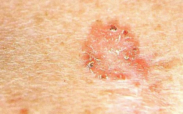
Bowen's disease is an intraepidermal cancer that tends to become squamous. It is an inflammatory disease of a chronic nature, which is associated with an excessive proliferation of atypical keratinocytes. Occurs in older people.
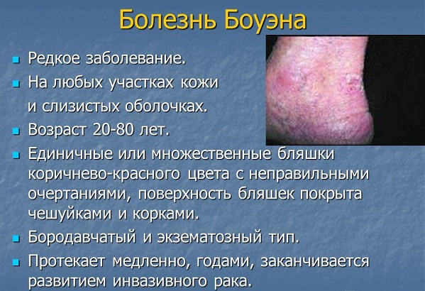
The tumor has invasive growth, grows not only in the epidermis, but also in deeper tissues. It can be located on any part of the skin and mucous membranes, but more often on the trunk.
Elements have the appearance of spots of pink shade with indistinct rounded edges. Under them there is an infiltrate, due to which the formations rise slightly. They are rough to the touch, covered with scales. When sloughing off the latter, an erosive, bleeding surface opens.
Paget's disease is a metastatic adenocarcinoma. The source of growth, as in Buen's disease, is located intraepidermally. The typical location is the mammary glands, most precisely the nipple zone and its areola.
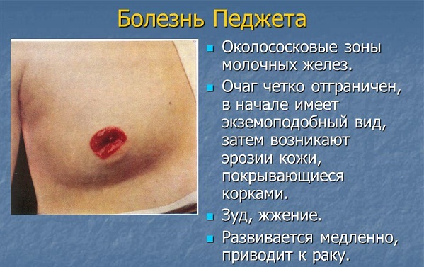
The growth of the tumor is infiltrating (it grows to the underlying tissues). Clinically manifests itself as a unilateral itchy plaque with clear contours in women over 40 years of age. The surface is covered with scales and crusts. The element increases in size, begins to metastasize. The outcome is breast cancer.
Diagnostics
Skin neoplasms, photos and descriptions of which were presented above, are diagnosed using general principles. These include the mandatory collection of anamnesis (patient complaints, clinical manifestations of the disease), examination of the patient, a thorough visual examination of the formations and the analysis of data from clinical and instrumental examination methods (MRI, radiography).

The main thing in the final diagnosis is the histological method. It is a microscopic examination of an area of pathologically altered tissue in order to identify atypical cells.
Treatment of neoplasms on the skin
Methods of treating skin neoplasms include medical, radiation and surgical. The latter is radical (i.e. allows you to get rid of the disease as completely as possible).
Skin neoplasms are medically treated with antibiotic therapy (photos and detailed descriptions of drugs can be found in clinical reference books), NSAIDs, opioid analgesics and hemostatic drugs. This method is used as a symptomatic treatment, it helps to alleviate the patient's condition, to somewhat improve the quality of life.
The surgical method is based on the elimination of the neoplasm. The goal of treatment is to finally get rid of the disease, prevent relapses.
Radiation therapy is more often performed for malignant processes, especially in cases where surgical resolution is impossible. Also, the goal is to prevent the resumption of tumor growth and its metastases.
Removal of skin lesions
Ways to remove skin lesions are:
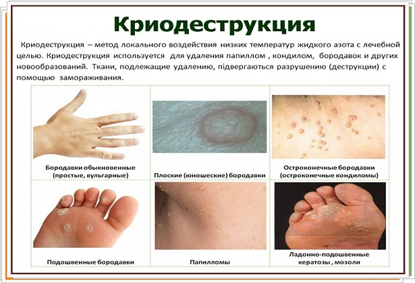
- Cryodestruction (with the help of liquid nitrogen, the tumor is frozen, later it dies and disappears). The method is not applied on the head, with large formations and with their intradermal localization.

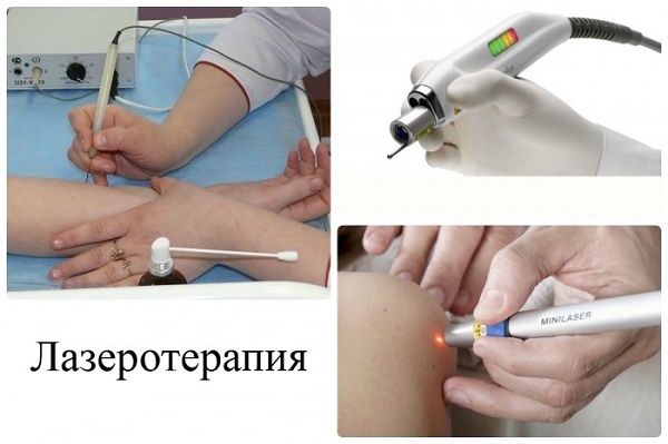
- Laser therapy. With the help of laser energy, pathologically overgrown tissues are "burned".

- Surgical excision. The scope of the operation can be different - removal of a tumor within healthy tissues (fibroma, nevi), together with a capsule (atheroma, lipoma), etc. up to excision of the surrounding skin, subcutaneous tissue, nearby lymph nodes (squamous cell carcinoma and other malignant formations).


In order not to miss the moment when new growths on the skin begin to threaten life, you need to understand what you have to deal with. Having studied the photos and descriptions of common pathologies, one can assume the source of the disease and contact a specialist in a timely manner.
A view of skin lesions
How to distinguish a dangerous mole from a safe one:
https://www.youtube.com/watch?v=4q_FgHF7-II
Characteristics of benign skin neoplasms:
I have a lot of moles (about 50). Who also, tell me, do you sunbathe in the sun?
I also have a lot of moles and the doctor recommended not to sunbathe at all. I don't know what to do, because I love to sunbathe.
Neoplasms on the skin (there are quite a few photos and descriptions of which in the article) are most often due to the human papillomavirus. But you should still be careful and in every case, urgently go to an appointment with a dermatologist.
"Also, the goal is to prevent the resumption of tumor growth and metastases." This is a quote from your opus. Do you even understand the absurdity of the construction of your proposals? According to your logic, it follows. that without prevention there are no relapses and only thanks to it they are renewed!
For a long time we will be "at war" with oncology thanks to such authors!
I re-read what you wrote several times, do you even understand how exactly you mastered this text? An unintelligible vskukorek about "fight". You need to go to the army, go and miss the war already.
a very useful article Especially people like me with a hereditary predisposition to cancer