Facial anatomy is a basic knowledge for cosmetologists. The skin is an equally important organ, like the stomach or liver - it protects the body from all sorts of environmental influences. And it is with the correct cosmetological effect on the skin of the face that you can not only remain attractive and young for many years, but also healthy - the better the skin condition, the stronger the immunity.
Anatomical features of the face
The face is a complex connection of muscles, blood vessels, nerves and veins. The internal structure, which is a rather complex and intricate mechanism.
In order to properly carry out aesthetic and medical procedures, one should take into account the complex of interrelated features of the skull, the placement of facial muscles, as well as their relationship with the lymphatic system, vascular network and the structure of the facial nerves.
Skull structure
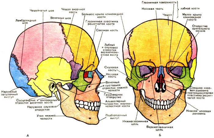
The human skull is the main protection for the facial muscles and nerves, which are responsible for the facial mobility of the face. In total, the skull contains 23 bones - that is, 8 paired and 7 unpaired. All of them are divided into 2 groups: facial and medullary bones.
Facial bones are smaller paired bones:
Nasal.
Palatine.
Zygomatic.
Lacrimal.
Upper jaw.
Inferior turbinate.
Unpaired facial bones:
Lattice.
Sublingual.
Coulter.
Lower jaw.
This group influences the normal functioning of the respiratory and digestive organs. The brain bones in total consist of paired and unpaired bones.
They are located above the facial region, form some parts of the face, namely:
Frontal tubercles.
Eye sockets.
Frontal area.
Whiskey.
The nasal cavity.
The paired bones are the parietal and temporal small bones, and the unpaired ones are the frontal, occipital and wedge-shaped. All parts of the skull are connected to each other with special "seams".
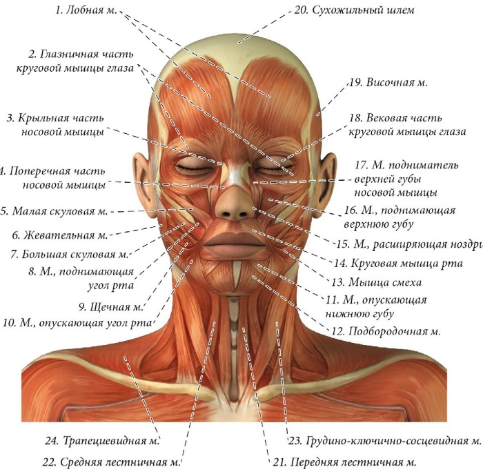
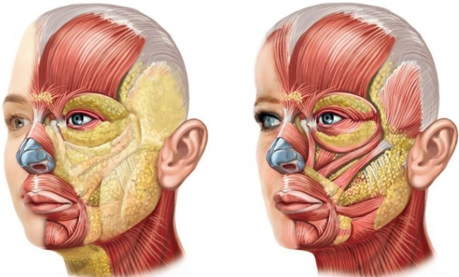
Facial anatomy for cosmetologists pays special attention to the muscle structure - soft tissues, which contract when a person is nervous.
According to myology, the science of muscles, it is possible to trace 1200 combinations of muscle work on the face, which reflect different states of emotions and well-being. Such facial expressions are possible only with the joint contraction of several muscle groups - different combinations of their work form on the face certain emotions of bliss, pain, disgust, interest or complacency.
Usually, most of the facial muscles are structured, with one end attached to the bone, and the other to the deep layer of the skin.
But on a person's face there is a group of 4 deep muscles, which are attached to the bone on both sides, and provide chewing actions:
Temporal.
Medial
Chewable.
Lateral pterygoid.
Aesthetic cosmetology does not work with such deep muscles, but their condition, tone and activity directly affect the condition of the facial skin and the shape of the oval.
Facial muscles are characterized by a thin form of structure from a flat fleshy part. They are mostly found in the subcutaneous tissue of the face. With this muscle contraction, several folds are formed, and they are located perpendicular to the corresponding fibers.
The main reason why changes in human facial expressions occur is the sensory effect of the nervous system on muscle work, which manifests itself in the corresponding sequential response of the muscles on the face.
The change in facial expression is due to the internal state and feelings of a person.
Such changes are possible with the help of 16 main muscle groups:
Muscle type
Functions
Occipital-frontal muscle
This muscle includes two paired smaller muscles. It tightens the skin of the forehead, keeps the eyebrow line. Due to the loss of muscle tone, over time, the eyebrows begin to drop and create sagging eyelids and age-related folds. With her vigorous activity, transverse folds appear - between the eyebrows and on the forehead.
Frontal muscle (upper part)
Controls facial expressions from the outside of the forehead to the tip of the eyebrow. During her activity, her forehead wrinkles around the entire perimeter.
Superciliary muscle
The small muscle that is responsible for the wrinkling of the forehead is located among the right and left frontal muscles, above the inner base of the eyebrows.
With its help, frowning, agitation or pain is expressed. Over time, this muscle provokes the appearance of vertical wrinkles on the forehead.
Circular muscle of the eye
Anatomically located along the perimeter of the eye. Consists of 3 parts that contract without affecting other parts of the muscle: the orbital, secular and lacrimal parts. Loss of their elasticity provokes the appearance of "crow's feet".
Muscle pyramidal (leg of the frontal muscle)
This muscle is located at the tip of the nose. When it moves, the top of the eyebrow stretches, which is why vertical folds are formed between them. Its other name is the muscle of the threat or proud.
Muscle above the upper lip
Allows you to wrinkle your nose, move your nostrils and lip tips.
Wing muscle of the nose
When it contracts, the facial expressions of the tip of the nose change, the nostrils expand.
Nasal (transverse) muscle
It covers the entire upper base of the nose, with its activity, mimic wrinkles appear in the form of swallows near the lips. The nasal muscle also pulls the skin of the cheeks with it.
Small zygomatic muscle
The base of the muscle is at the top of the cheekbones, and stretches towards the soft tissues at the corners of the lips. The lips react to her work, they can rise by 1 cm, and with this movement they create a nasolabial furrow.
Large muscle or muscle of laughter
Its beginning is in the back of the zygomatic bone, and the end is in the deep skin tissues near the mouth. When it moves, nasolabial folds appear. Which, in turn, press on the cheeks, because of which they bulge slightly and rise. With this movement, the cheeks provoke the appearance of wrinkles near the eyes.
Buccal muscle
When it contracts, the cheeks are inflated. This is the "safest" muscle, it does not provoke the appearance of facial wrinkles.
Muscle that lifts the corners of the mouth
Its base is in the front of the upper jaw, under the eye, and this muscle ends in the deep tissues above the lip. Due to the fact that it is poorly developed, its reduction can be noticed only during strong aggression.
Circular muscle around the labial line
The flat muscle has the shape of a circle, which consists of two semicircles: upper and lower. They connect near the lips. These muscles begin to move when eating or talking.
Muscle of the corner of the mouth (triangular)
Located near the chin muscle, its beginning is attached to the lower jaw, and the end is near the skin near the corners of the lips. Its reduction greatly affects the facial expressions - the corners of the lips eventually lower and curve the lip line.
Chin muscle or muscle fiber bundle
Located deep under the skin of the chin. During its contraction, the lower lip rises, which causes bumps on the chin.
Subcutaneous muscle of the neck
Refers to the facial muscles of the facial group - when this muscle moves, then almost all the muscles on the face react.
The golden rule of all cosmetic procedures is to follow the massage lines.
It is very important for cosmetologists to know the anatomy of facial massage lines
This ensures the tone and elasticity of the muscles that support the face frame, guaranteeing youthful skin. Cosmetologists recommend adhering to the massage lines scheme, since they are areas that are least prone to stretching of skin tissues.
If you regularly maintain the tone of the facial muscles and gently, along the appropriate massage lines, massage, you can tighten the shape and create a more expressive outline of the oval features.
All muscles, during their contraction, change facial features, expresses the internal state of a person. Since each muscle is associated with a certain state of mind, which is displayed on the face in the form of a change in its shape, a corresponding facial expression occurs, as a result of which wrinkles and folds appear over time.
Facial anatomy for cosmetologists focuses on the important role of the normal functioning of the lymphatic system on the condition of the skin.
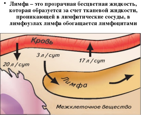
This system is a very dense capillary network that is present in all organs and tissues of the body. Disruption of the lymphatic system often affects the condition of the skin of the body - it loses its beautiful color, elasticity and velvety. The loss of these qualities due to problems of lymph flow in twins is noticeable in the condition of the facial skin.
The lymphatic system refers to the vascular system of the body. Under its influence, lymph moves in the body, a transparent liquid, which, like blood, circulates throughout the human body.
But the lymphatic system does not have a pump, the function of which in the circulatory system is performed by the heart, and therefore the movement of lymph occurs very slowly - towards the large veins, at a speed of 0.3 mm / s... Therefore, it is always worthwhile to activate its work by mechanical action - massages, baths and cosmetic procedures - such manipulations will speed up the work of the glands.
This system cleanses the body.
Important functions of the lymphatic system are:
The distribution of fluid in the body.
Transport of nutrients from tissues.
Protecting the body from bacteria, supporting the immune system.
It consists of:
Vessels.
Nodes.
Duct.
Tonsils, thymus.
In the human skull, the lymphatic system has 7 groups of nodes:
Occipital.
Cervical.
Behind the ear.
Cheek.
Submandibular, are in the chin triangle.
Parotid.
Chin.
Therefore, if the lymphatic vessels are clogged, and the work of the system is disrupted, many diseases arise on the skin, which can manifest themselves in the form of acne, boils, and other rashes.
If lymphatic drainage procedures are carried out regularly, then these manipulations will have a good effect on metabolic processes in the tissues of the body. So, for example, you can reduce the swelling of the face, improve its contours and elasticity, normalize the tone of facial muscles with regular massage. It is very important for a beautician to know the direction of lymph flow on the face.
Since it is a complex network of capillaries, lymph flow has several directions:
AND) The lymph that flows through the tissues of the face enters here with the help of superficial vessels. The lymph flow corresponds to the blood veins.
Superficial lymphatic vessels are grouped into anterior and posterior:
Posterior vessels supply lymph to the back of the head. There they pass into another group of vessels - the occipital.
Anterior vessels located simultaneously from the forehead, eyelid, crown and temples. These vessels are connected to nodes near the ears, through which lymph continues to move through the vessels down the neck.
B) From the eyelids, from the nose, cheeks and lips, the lymphatic network begins, its movement is partially directed to the submandibular triangle, the submandibular nodes are located there. Another part of these vessels interrupts their circulation in the buccal nodes.
IN) The chin lymph nodes, which are located under the hyoid bone, are supplied with lymph from the vessels near the lips and chin.
D) Deep vessels from the hard and soft palate direct their lymph flow to the deep nodes of the parotid gland.
Skin on the face
https://www.youtube.com/watch?v=0z8cU4gOfyw
The skin of the face performs the protective function of the body from the external environment. In order for this protection to take place in the best possible way, cosmetologists in every possible way maintain the normal state of the skin of the face, because flabbiness, wrinkles, rashes or dryness are not only ugly aesthetically, but also signs of deterioration in the motility of cell metabolism, or malfunctioning of skin tissues.
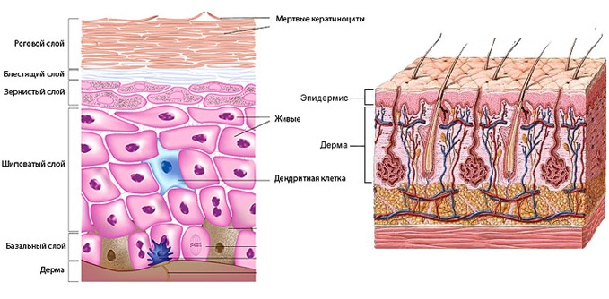
Facial anatomy for cosmetologists describes in detail the structure of the facial skin, which consists of many cells, and their healthy state affects the appearance of a person.
The vital activity of cells is very similar to the life of all creatures - they absorb oxygen, feed, and have the ability to reproduce. Although cells are the smallest living units, they contain a large number of organelles and elements that ensure the normal life cycle of each cell, and respectively - its owner:
Ribosomes provide protein synthesis in the cell.
The centrosome takes part in the regeneration of nutrients.
Lysosomes are responsible for the metabolism and absorption of nutrients.
Cytoplasm - retains the activity of all nutrients in the cell, except for the nucleus.
Microvilli are responsible for the transport of substances from the cell through the membrane.
Kernel - stores information about hereditary traits.
The epidermis is the first upper layer of the skin of the face, it serves as the main barrier of protection, responsible for getting a tan when exposed to sunlight. Almost all cosmetic procedures are aimed precisely at maintaining the elasticity and tone of this particular layer of the skin. The epidermis in its structure, has several layers of cells - lower, prickly, granular, flattering and horny.
The last layer of the skin, the stratum corneum, is the uppermost one and consists of dozens of corneocytes - cells that are the most mature on the face, and therefore any metabolic processes stop in them. These cells are already old, and therefore contain little water, keratin and do not have nuclei.
Their main function is to create a protective barrier against external factors for the skin of the face. Usually, within 28 days, old cells are peeled off, and new ones grow in their place - here there is a constant process of new cells emerging and old ones peeling off. Most mechanical and chemical peels work at this level. The second layer of the facial skin is the dermis.
It consists of two levels:
Mesh layer - the level at which the networks of lymphatic and blood vessels, hair follicles, sebaceous glands and all fibers are located - they are responsible for the smoothness of the skin.
Papillary layer concentrates nerve endings, outgrowths and capillaries.
You can do any procedures on this layer of the skin with the help of deep-dropping agents with active compounds. Most cosmetics are superficial agents, so only a special education will help you choose the composition of the products that will penetrate the epidermis to the dermis.
The dermis is responsible for the production of elastin and collagen in skin cells. Therefore, when deep wrinkles appear, there is an immediate need to influence this layer of the skin, to ensure its elasticity, and to strengthen it.
The third, deepest, layer - subcutaneous fatty tissue, is responsible for storing nutrientsthat directly affect the condition of the skin. This layer of skin consists of many nerve and blood vessels, as well as fatty accumulations. The need to act on this layer of the skin arises with vitamin deficiency, when the face loses its healthy color.
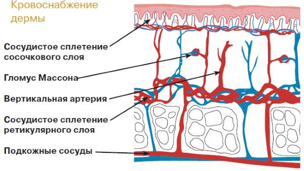
Vascular and nervous tissue of the face
Facial anatomy necessarily teaches the location of the vascular network on the human face - the small venous ducts that supply the facial tissues with important nutrients. For cosmetologists, the problem of blood vessels, or rosacea, is the most common complaint for which women turn to aesthetic medicine for help.
Couperosis is a genetic predisposition of almost every person to the manifestation of redness and irregularities on the skin of the face. But for all this property of the skin has different forms, and may be more or less noticeable.
The first signs of "asterisks", "streaks" can appear even in childhood, and only competent treatment and maintenance of vascular health can save you from aggravating the problem. If a girl has such a predisposition, then there is a possibility that the couperose mesh after 30 years will become very noticeable.
Treatment of rosacea of the facial skin requires a systematic approach - you need to regularly add aromatic oils to your daily care - this will strengthen the walls of blood vessels and prevent their potential damage, for example, during stressful situations.
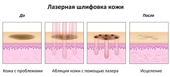
If the problem of rosacea already has a more pronounced condition, then the treatment procedure requires the use of hardware cosmetology:
Photorejuvenation - This is the most popular method as it has no age restrictions. This procedure takes place under the influence of impulses, increases the regeneration of the facial skin, strengthens it. Subsequently, this makes the vascular network on the face invisible.
Mesotherapy - the procedure provides the supply of skin cells with a concentrated complex of useful substances, due to which minor defects, such as rosacea, disappear.
Electrocoagulation - a procedure for removing large stars using an electric current.
Laser procedure will help remove pronounced vascular network.
Ozone therapy it is carried out only at the advanced stage of rosacea - during the procedure, damaged capillaries are removed under the influence of ozone oxygen using a microneedle.
An important knowledge in aesthetic cosmetology is also the structure of the nervous tissue - ectodermal formation from nerve cells, neurons. Its main task is the excitability and conductivity of nerve receptors and impulses from a particular organ to the central nervous system. They form a network of nerve nodes that perceive any irritation upon contact with them.
If the vascular or nervous system is damaged during the procedure, it is possible to break the symmetry of the face or provoke a pinched muscle or nerve.
Knowledge of the location of the vascular and neural networks on the face is a very important skill for a cosmetologist - when performing any injection technique, it is necessary to clearly understand where the lines of large vessels and nerve tissues pass in order to avoid further dangerous manipulations in these zones.
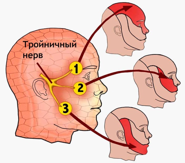
One of the important points of facial anatomy is the structure of the facial nerves - an unsuccessful procedure can cause a certain form of deformation or asymmetry of the face after the procedure. Together with muscles, facial nerves are responsible for facial expressions, and often it is nerve disease that can provoke facial distortion.
Facial anatomy for cosmetologists describes the structure of the facial nerve as one of the most difficult topics to study, since its scheme is very confusing - the facial nerve is 7 of the 12 cranial nerves that affect the activity of the facial muscles of the face.
Its complex topography is explained not only by the extension of this nerve through the facial canal from the temporal bone, but also by the constant complication of its circuit due to permanent processes in other directions:
The nerve itself is made up of fibers that run from several nuclei: motor fibers, sensory fibers, and secretory fibers. Then it penetrates into the opening of the ear canal.
From the parotid gland, 4 branches of nerves begin: the posterior auricular nerve, stylohyoid, digastric and lingual.
Another 5 branches depart from the parotid salivary gland: the temporal, zygomatic, buccal branches, the marginal branch of the lower jaw and the cervical.
The anatomy of the facial nerve is an intricate system of small canals in the face that send signaling responses to specific parts of the head or neck. The facial nerve is mainly responsible for the motor function of the muscles in the face.
Knowledge of the functions of each branch of this nerve is very important for cosmetologists - this is the only way to determine the main problem of impaired sensitivity and facial expressions, and determine the subsequent treatment tactics.
Thank you so much. Very informative material. But, since I have no medical education, I would like to read about the function of each branch of the nerve. Please tell me the literature. Thanks again.

 Don't miss the most popular column article: Fashionable haircut for short hair. Photo, front and back views.


Thank you so much. Very informative material. But, since I have no medical education, I would like to read about the function of each branch of the nerve. Please tell me the literature. Thanks again.